How do you see?

Have you ever wondered how you see?

I’m naturally curious, so if you’re anything like me, you have probably wondered at some point just how the phenomenon of sight occurs.

(If you’re not, maybe you should start reading now..)

Thankfully, during the course of optometry school, this process was mildly engrained in us, and today, I’m excited to share it with you.

Vision: Explained

First things first: light.
Everything that you see in everyday life corresponds with an electromagnetic frequency within the visible spectrum of light.
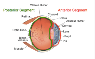
Light enters the eye through the clear cornea, passing through the lens, and ends up at the retina.  Together, the cornea and the lens focus the light to attempt to provide a distinct pattern of light on the retina.  When they aren’t enough, spectacle or contact lenses are added to help focus the light.
When the light hits the retina, it is sensed by special cells called photoreceptors.  The role of photoreceptors is to take the light and transform it into an electrical signal.
Within your eyes, there are different types of photoreceptors: three that require more light to function, and so are utilized in brighter lighting  (cones), and one that requires little light to function, allowing it to work in darker conditions (rods). Each of these have specific wavelengths that they are more sensitive to.   Three different cells with three different specific wavelengths provides for the perception of color in brighter lighting conditions.
Additionally, cones and rods have specific functions.  Cones are responsible for your best vision.  For this reason, they are highly concentrated in the most central part of your vision to allow you to see clearly when looking straight ahead.  Rods, on the other hand, provide less precise vision, and are located further out from the very central point of vision.
The signal produced by the photoreceptors is then transmitted through additional cells in the retina. These cells help to group, enhance, and refine the signal as it makes its way to the retinal ganglion cells that then pass out of the retina as the optic nerve.
The optic nerve (well, after some minor-ish detours) then passes to the first visual center in the brain – the lateral geniculate nucleus.  While it would seem like a primary place for image processing, as this structure is comprised of 6 layers with divided input based on specific types of retinal ganglion cells, relatively little processing occurs here.  Rather, it would seem that the lateral geniculate nucleus simply serves to help combine information from both eyes before going further in the brain, or provides an ideal midpoint for information modulation.
From the lateral geniculate nucleus, the signal passes to the primary visual cortex (by way of optic radiations).  This is where the first visual processing occurs, allowing for the beginnings of depth perception, a determination of object orientation, and detection of movement.
The signal continues to pass though the other 5 layers of the visual cortex, becoming increasingly refined along the way, until full perception of the image is achieved!
However, simply creating an image does not allow you to react to what you see.  Rather, once fully processed in the visual cortex, signals are sent to other areas of the brain (such as Brodmann and Wernicke’s areas) that then give context to the image.  Once context is achieved, signals are set to respond appropriately in some form of action – be it smiling at a funny picture, stepping out of the way to avoid someone walking towards you, or reading words off a page!
If you learned something from this post, please share it with a friend or family member!  If you liked it, please subscribe, or like my page on Facebook! And as always, if you have any questions or comments, please contact me – I’d love to hear from you!

Virtual Learning Tips and Tricks

Happy Monday, y’all!

Ready or not, a new school year is upon us.  However, for many, it looks very different than years in the past, as virtual schooling has become a reality for children of all ages across the country.  Children from Kindergarten through high school (and even through college)  are now needing to sit still at home in front of computer screens for hours at a time, completing virtual instruction.
Unfortunately, this mode of education poses significant problems for the visual system.
In a previous series, I discussed Computer Vision Syndrome in adults.  However, this condition is not limited by age.  Rather, as screen time increases, children are reporting  increasing symptoms of Computer Vision Syndrome as well.
As a review from before, some common symptoms of computer vision syndrome are:
  • Headaches (especially around the eyes)
  • Eye strain
  • Eye fatigue
  • Double vision
  • Blurry vision
  • Dry eyes
But in an environment where virtual learning at times seems to be the only viable option, what can be done to prevent these symptoms in children?
A Facebook group that I am a part of (VTODs on Facebook) has compiled some tips and tricks for optimal ocular health in this new situation, and so, for today, I’d like to share some of them with you!
  1. Blue light filters: Blue light filters block blue light (which is emitted from screens), which has been shown to have effects on circadian rhythms and potentially cause ocular fatigue secondary to the screen flicker rates.  If you’re ordering new specs for your child, consider adding a blue blocking filter.  If your child doesn’t wear glasses, blue light filters are sold as prescription-less glasses, as well as filters that can be put on screens!
  2. Reading glasses: When using screens for long periods of time, it may be beneficial for your child to use low powered reading glasses to relax how hard they have to focus their eyes to clearly view the screen.  Low plus (+1.25-+1.50) lenses can be bought over the counter at most stores.
  3. 20/20/20 Rule:  As with adult Computer Vision Syndrome, encourage your child to look at something 20 feet away for 20 seconds every 20 minutes to give their eyes a break!
  4. Distance: Make sure all screens are at least an arms length away from your child!
  5. TV: Consider casting your child’s virtual class to a TV screen for easier viewing.
  6. Activities: Try to have your child do some form of physical activity (jumping jacks, jump rope, running, etc) for at least 5 minutes to use some energy before having to sit still
  7. Windows:  If possible, have your child’s school set up be next to a window so that they can easily look far away.
  8. Go outside:  During any breaks, go outside!  This helps relax eyes, and is good for overall health!
If you learned something from this post, please share it with a friend or family member!  If you liked it, please subscribe, or like my page on Facebook! And as always, if you have any questions or comments, please contact me – I’d love to hear from you!

(Neuro) Optometry in Focus: Strokes

So, as I discussed in my Introduction: Hannah Vollmer, OD post, one of my greatest optometric passions is neuro-optometric rehabilitation, which is the field that I completed a year long residency in.  During my residency, I had the opportunity to work in an in-patient rehabilitation hospital, which was one of the most rewarding experiences in my career to date.

As a neuro-optometry resident, I worked with a significant number of people who were in the process of recovering from strokes. Prior to residency, I honestly had little understanding of the effects of strokes or the potential treatment strategies to be implemented in stroke survivors, despite having four years of graduate level, medically based education and a positive family history for the condition.

Maybe I lived under a rock, but I’m guessing I’m not alone.

Over the past year, I have learned a lot about working with stroke survivors, but I’m still nowhere near an expert.  Nevertheless, in today’s post, I’ll try to share a few things that I’ve learned over the past year, especially regarding how strokes impact visually related tasks.

Enjoy!

Strokes: In Focus

What is a Stroke?

I suppose talking about what a stroke is would be a good first step…

Medically, strokes are called cerebrovascular accidents, or CVAs.  This term does a good job at explaining what a stroke is: a condition in the brain (cerebro) that occurs as a result of problems with blood flow (vascular).

Strokes can be either ischemic or hemorrhagic.

Ischemic strokes occur secondary to a blockage that prevents blood from entering an area of the brain.  This is the most common type of stroke, accounting for nearly 90% of all strokes.

Hemorrhagic strokes, on the other hand, occur when a blood vessel breaks or leaks within the brain.  These are relatively rare, and are most often associated with aneurysms (essentially an outpocketing of the blood vessel) or very high blood pressure.

Who Gets Strokes?

According to the CDC, approximately 795,000 people in the US alone have a stroke each year.  This equates to one person every about 40 seconds.

Out of these, approximately 140,000 people die (1 every 4 minutes).

Around 610,000 are first time strokes.  The other 185,000 are a recurrence.

Crazy, right?

Strokes are more common in patients with vascular problems, such as high blood pressure, high cholesterol, and diabetes.  Higher risk is also associated with smoking and obesity.

While strokes are often associated with older age, over 1/3 of the people who are hospitalized with strokes are under the age of 65.

Takeaway: Anyone can have a stroke at any time.  However, certain conditions or behaviors significantly increase the risk of having a stroke!

Symptoms of a Stroke

Considering how little information I hear about strokes on a regular basis, I feel like this is one aspect that the public health community has made huge strides in – knowing the symptoms of strokes.

Remember the acronym? FAST?

For a review, it stands for:

  • Facial droop
  • Arm weakness
  • Slurred speech
  • Time to act

These are some of the most common symptoms associated with a stroke.  However, it’s important to know that these can also occur as part of what’s called a transient ischemic attack, or TIA.  In these cases, as the name implies, the symptoms are transient, or short lived.  Just because the symptoms are not permanent, though, does not negate the seriousness of the presentation.  TIAs are considered a medical emergency, as they often precede a full-scale stroke.

Despite the widespread knowledge of the FAST acronym, these symptoms fail to include any visual symptoms that may precede or accompany a stroke.  I guess that’s where I come in…

Visual Symptoms of a Stroke

First things first: strokes may often be preceded by visual TIAs.

These visual phenomena can be described in a number of different ways, such as:

  • Vision loss (typically in one eye)
  • Double vision
  • Dizziness/sensation of world moving
  • Loss of part of the visual field

As with all TIAs, these symptoms are relatively short lived, normally lasting under an hour, but are still suggestive of an impending full-scale stroke, with the greatest risk being in the first 24 hours after the event.

While these three symptoms may precede a stroke as visual TIAs, they may also present consistently, either before or with a stroke.
For instance, sudden vision loss in one eye may be a result of occlusion (blockage) of the central retinal artery, or a branch of the central retinal artery (CRAO or BRAO).  These conditions are both considered a stroke of the eye and require immediate medical attention due to the strong association between these conditions and risk of large-scale stroke.
New onset, persistent double vision, from my experience, is more likely to occur in conjunction with the full ischemic or hemorrhagic event.  This annoying symptom presents secondary to damage to the neural pathways responsible for controlling the four cranial nerves that innervate the six extraocular muscles.  With improper innervation, the musculature becomes imbalanced, changing the natural position of gaze for one eye, creating the symptom of double vision.  This eye turn may or may not be easily observable, depending on the direction and degree of turn.

Dizziness, or a sensation of the world shaking or moving (oscillopsia), again may present as a symptom of the stroke itself.  Oscillopsia occurs as a result of damage to a part of the brain that normally inhibits movement of the eyes (if you inhibit an inhibitor, you get movement).  Visually, this presents as nystagmus, or a shaking of the eyes.  The direction that the eyes move is directly determined by the location of the stroke itself.  This is one of the most frustrating visual symptoms for patients who have had strokes, as it not only reduces vision (because the eyes never sit still long enough to fully focus on an object), but also causes dizziness (frequently with nausea and vomiting) and disorientation.

Loss of part of the visual field is another significant visual sign associated with strokes.  This symptom may occur secondary to damage to a number of areas in the visual pathway – whether it be in a lobe of the brain that the nerve fibers traveling from the eye to the visual cortex pass through, or in the visual cortex itself – with symptoms varying by the specific location affected.  Interestingly, due to the crossing of nerve fibers from each eye relatively early in the ocular pathway, visual field deficits generally present as loss on the right or left halves of both eyes.  This is referred to as a homonymous hemianopsia – homonymous meaning same, hemianopsia meaning half of the visual field.  Due to the oddly reversed nature of physical placement of retinal nerve fibers and the corresponding visual field, a stroke on the left side of the brain will be associated with right sided visual loss, and vice versa.

What to Do?

If you, or someone you know, experiences any of these symptoms, immediate action is imperative!  When you’re working with brain tissue, time is money.  Or, in this case, life.  The sooner an impending, or even large-scale stroke is caught, the better the outcome.  If caught soon enough (under 5 hours from onset), a medication may be administered to get rid of the blockage, potentially reducing or completely reversing the effects of the stroke.

However, unfortunately, simply going to the hospital isn’t always enough, as not all stroke centers are created equal (though, if you only have the option of hospital or no hospital – get to the hospital).  Optimal treatment would be at a comprehensive stroke center, which is required to have:

  • The ability to treat ALL types of strokes
  • 24/7 access to minimally invasive procedures to treat stroke
  • Neuroscience ICU
  • Neurosurgery

Treatment after a stroke varies widely by symptoms and their severity.  For the sake of time, though, I’ll discuss stroke rehabilitation (specifically the role of optometrists in stroke rehab) in a later post.  Stay tuned!

If you learned something from this post, please share it with a friend or family member!  If you liked it, please subscribe, or like my page on Facebook! And as always, if you have any questions or comments, please contact me – I’d love to hear from you!

Optometry in Focus: Floaters

How many of you have floaters in your vision?

You know – those little (or sometimes bigger) black spots, dots, and squiggles that course through your vision that normally become more noticeable when you’re looking at a white/bright background?

Have you ever wondered what they are (other than annoying) and why you have them?

Let’s chat!

Floaters: In Focus

Ocular Anatomy

As normal, before we can fully dive into what floaters are, there needs to be a little bit of context – namely, knowledge of some ocular anatomy.

From those who have been following these posts, in my article “Why Does My Eye Doctor… Part 3” where I discuss the reason for the air puff test, I talk quite a bit about the anterior chamber (and segment) and the liquid, or aqueous, that fills it.  For today, however, we’re going to be hopping further back in the eye, and going behind the lens to the posterior segment (highlighted in green in the picture below).

Function and Structure of the Eye: Vision and Posterior Segment - Medicine  Gemp2 with Gemp2 at University of Witwatersrand - StudyBlue  

This back portion of the eye is made of several components (retina, optic nerve, choroid, other vasculature), but today’s focus is primarily on one: the vitreous.

Vitreous, or vitreous humor, is a jelly-like substance that functions to absorb shock, maintain the shape of the eye, keep the retina intact, and transfer molecules (like oxygen) from the front to the back of the eye.

In youth (generally), this jelly-like substance has a pretty uniform consistency – the fibers that make it up are evenly spaced, with water-loving molecules spread throughout to maintain proper water content and spacing.

However, with time, this spacing changes.  The fibers that were once perfectly spaced begin to clump together, as the water component of the vitreous increases.  These fiber clumps cast shadows on your retina as light comes in, causing you to see floaters!

Types of Floaters

While simple, normal, changes in the vitreous are the most common type/cause of floaters, there are others.  For instance:

  • In cases of ocular inflammation, there can be cells (specifically white blood cells) that end up in the vitreous.  This is commonly referred to as vitritis, and may present with complaints of floaters.
  • Sometimes when there is bleeding around the retina, the blood can escape into the vitreous, causing a vitreous hemorrhage.  Patients at times report seeing red floaters in this situation.
  • As the vitreous begins to clump together, it can put tension on the retina.  Most of the time, this is focused around the optic nerve, where the vitreous is strongly attached.  Eventually, the vitreous pulls enough and detaches, forming a c-shaped, or spiderweb-like floater that’s relatively close to the center of vision.  This detaching of the vitreous from the retina is called a posterior vitreous detachment, and though it sounds really bad, it’s perfectly normal!
  • In some cases, in conjunction with the vitreous pulling on the retina, a piece of the retina may actually tear off, forming either an operculated retinal hole, or a retinal tear (which then may lead to a retinal detachment).

Who gets floaters?

The short of it is – everyone!  However, the number of floaters and the time of onset varies dramatically from person to person.  As an example: I’d like to think I’m pretty young, but I have a lot of floaters – so many that I’ve been known to question if I’m seeing my floaters or my patients floaters during an exam, as well as wonder if I’m seeing bugs flying around or just some new floaters!  My dad, on the other hand, says he rarely if ever notices floaters in his vision!

When to Be Concerned

While floaters are very common, sudden changes in floaters can indicate a problem (such as inflammation, bleeding, or detachment), in the back of the eye.  For this reason, anyone who experiences a sudden increase in flashes and floaters should immediately contact their eye care provider!

If you learned something from this post, please share it with a friend or family member!  If you liked it, please subscribe, or like my page on Facebook! And as always, if you have any questions or comments, please contact me – I’d love to hear from you!

Fun Post Friday: Fact Checker – Carrots?

Happy Friday, guys!

First things first: After seeing the response to last week’s Fun Post Friday, and realizing how nice it is to end the work week with something less mentally taxing, I’ve officially decided to make Fun Post Friday’s a thing!

With that in mind, I honestly don’t know yet what all these posts will look like – I’ll just try to keep them fun, probably a bit shorter, but still informative.

And with that, let’s dive in to this week’s post!

Since it’s political season… again… (does that season ever end?), it only feels fitting to write a fact checker post about a common claim involving your eyes – carrots!

Claim: Eating Carrots Improves Vision

I’m firmly convinced that this is a parent’s favorite way of convincing their child to eat veggies – especially carrots.  I mean, who wouldn’t want to have bionic vision from eating healthy foods?!

Unfortunately, for all the hopeful parents and children out there, this notion is (almost) completely false!

So, where did it come from?  Supposedly, this myth originated in WWII, after the British Royal Air Force developed a new type of radar technology that allowed them to shoot down German planes at night.  Rather than divulging their newfound technological advancement to the public (which would not be particularly advantageous for *ahem* winning the war…), they reported that the pilots’ “incredible night vision” was due to carrots.

Interestingly enough, however, vitamin A, one of the primary vitamins in carrots, is actually vital for night vision, as it is a precursor molecule for the photopigment rhodopsin.  This pigment is responsible for absorbing light, as part of the phototransduction cascade, in which light is converted to a neurological signal that is then transmitted to the brain.

Additionally, vitamin A is important in maintaining the health of the front surface of your eye.

Hold up.
Now where’s the fact checker – this post is literally disagreeing with itself – right?

Not so fast.
While vitamin A is important for vision, increased consumption will only improve vision in the case of vitamin A deficiencies, which are rare in the United States and other first world countries.  Additionally, excess vitamin A intake can actually be detrimental to your health.

Vitamin A toxicity is a condition that can present either either acutely or chronically, depending on the extent and duration of excess vitamin A intake.  In the acute form, common signs and symptoms are:

  • Drowsiness
  • Irritability
  • Nausea
  • Vomiting

 which may or may not be associated with increased intracranial pressure.

Chronic vitamin A toxicity more commonly presents with:

  • Hair and skin changes
  • Headache
  • Weakness
  • Fractures
  • Pseudotumor cerebri

In both of these, the most concerning presentation is often observable ocularly: increased intracranial pressure/pseudotumor cerebri.

As the name implies, vitamin A can cause increase in the fluid pressure inside the skull.  Since this is a confined space, the increased pressure acts somewhat like a tumor (hence pseudo, or false, tumor). With nowhere else to go, the increased fluid is transmitted through the optic nerve, disrupting the normal layers, and presenting as swelling, or papilledema (by definition swelling of both optic nerves secondary to an increase in intracranial pressure).

Unfortuantely, this swelling of the optic nerve causes compression of the nerve fibers, and, if left untreated, may result in permanent vision loss.

Now, granted, it can be difficult to consume enough carrots to create this type of scenario.  Vitamin A toxicity is more often associated with vitamin A supplement use.  More commonly, an excessive carrot consumption will only turn your skin – specifically in the palms of your hands and soles of your feet – orange (caretenosis).  However (not from personal experience or anything), eating around a pound of carrots/day for three months is a really bad idea.

Takeaway: Vitamin A, which is found in carrots is necessary for vision, but will only improve vision in rare cases of deficiency.  Excess vitamin A intake can result in vitamin A toxicity, which, if left untreated, may cause permanent vision loss.

And there you have it!

If you learned something from this post, please share it with a friend or family member!  If you liked it, please subscribe, or like my page on Facebook! And as always, if you have any questions or comments, please contact me – I’d love to hear from you!

Why Does My Eye Doctor… Part 4

Welcome back to my “Why Does My Eye Doctor…” series!

Have you ever wondered, “Why does my optometrist care so much about my medical history? That has nothing to do with my eyes!!”

If so, you’re not alone.  It can definitely seem weird being asked (or being the doctor and asking) about general medical history while at the eye doctor, but I promise it isn’t just to be nose-y!

Believe it or not, your ocular health is closely related to your systemic health, and your eyes are often one of the first places to show systemic disease!

Sound crazy?  Let’s take a look at four (relatively) common conditions that can be seen in your eyes!

Diabetes

  • Diabetes is a disease that affects the small blood vessels in your body.  (Okay, it affects larger vessels too, but for the sake of the eye, we’re focusing on the smaller ones.)
  • In the eyes (specifically the retina), this can cause bleeding, or hemorrhages, as well as leakage of other materials from the blood vessels.  If this bleeding or leakage happens to be around the macula, or part of the eye responsible for your best vision, it can cause swelling that then distorts your vision.
  • Fluctuations in blood sugar levels can also affect your vision, as they cause changes within the lens of your eye.  If you notice frequent changes in vision, it may be a sign of diabetes.

Hypertension

  • High blood pressure is, once again, a disease of the small blood vessels in your body.
  • The retinal disease (retinopathy) associated with hypertension occurs in 3 main stages.
    • Vasoconstrictive: This is a fancy way of saying that the blood vessels constrict in response to the increase in blood pressure.  The goal of the constriction (which is automatically controlled) is to decrease the amount of blood flow so that the normal vessel system of the retina isn’t overwhelmed.  This early stage is seen in the eye as arteriolar narrowing.  *I would include a picture, but it can be pretty hard to appreciate*
    • Sclerotic: The term sclerosis simply means hardening.  The constriction of the blood vessels in the vasoconstrictive phase occurs via a muscular layer in the arteries.  Like any muscle that is frequently used, over time, this muscular layer gets bigger, making the arteries harder.  As the arteries harden and become heavier, they compress the underlying veins.  This causes a characteristic appearance of blood vessel “nicking” as seen below, where the vein becomes diminished upon artery crossing.
Arteriovenous nicking - Wikipedia
    • Exudative: Once enough compression has occurred in the sclerotic phase, blood, fats, and additional components begin to leak out of the vessels, signaling a transition to the exudative phase.  In this stage, hemorrhages (areas of bleeding), exudates (hard, fatty deposits), and edema (swelling from leaking fluid) are common.  As blood vessel compression also reduces blood flow (stasis), clots are more likely to form, occluding, or blocking the vessel.  Without proper blood flow, ischemia (literally lack of blood flow) occurs, causing subsequent damage to affected tissues. 
This is Grade 3 hypertensive retinopathy. Diastolic pressure is usually  110-115 mmHg and retinal arteries lose their abilit… | Retinal photography,  Eyes, Eye health

Thyroid Disease

  • First things first: thyroid problems can come in two forms – overactive (hyperthyroid) or underactive (hypothyroid).
  • Both versions can be caused by autoimmune states.  The one most commonly associated with hyperthyroidism is Grave’s Disease, while the one most commonly associated with hypothyroidism is Hashimoto’s Disease.
  • In addition to systemic manifestations, both hyper and hypothyroidism may present in the eyes. Possible symptoms include:
    • Redness
    • Dryness and irritation
    • Swelling around the eyes
    • Protrusion of the eyes
    • Pain with eye movement
    • Inability to fully close the eyes
    • Double vision
    • Loss of vision
  • These symptoms may occur in existing cases of autoimmune related thyroid disease, or may be among the first presentations of the condition.  If your doctor suspects thyroid disease secondary to ocular presentations, they will likely ask a number of systemic questions that seem totally unrelated such as:
    • Unintentional/unexplained weight gain or weight loss
    • Sensitivity to heat or cold
    • Changes in energy levels (either very high or very low)
    • Changes in mood (such as being highly irritable or more depressed)
    • Changes in bowel movements
    • Changes in menstruation
  • If your doctor asks these seemingly off the wall questions, don’t freak out!  They’re trying to narrow down the potential causes of your symptoms so that they can send you to the right place to get the right diagnosis!

Multiple Sclerosis

  • While we’re on the topic of autoimmune diseases, it’s important to discuss Multiple Sclerosis.
  • In this condition, antibodies attack the myelin (protective coating) that surrounds nerves in your central nervous system (brain and spine).  This causes demyelination (literally, loss of myelin) around the nerve, which impedes transfer of the signal from the brain to its intended recipient.
  • One of the more common, and often early, presentations of multiple sclerosis is optic neuritis, or inflammation of the optic nerve.
    • Optic neuritis generally presents as mild-moderate vision loss in a single eye that comes on suddenly and may worsen for around 2 weeks, after which symptoms begin to improve.  Colors may seem less vibrant out of this eye, and there is often pain with eye movement.
    • As with any sudden decrease/loss of vision, it is important to see an eye doctor with these symptoms.
    • Looking in the back of your eye, the doctor will often see nothing out of the ordinary.  This is because the inflammation is behind your eye (retrobulbar).
    • Your doctor should order an MRI, with and without contrast, to aid in diagnosis.  The results of this test provide important information regarding future prognosis as:
      • 1/4 people with no other lesions on MRI have a diagnosis of Multiple Sclerosis within 15 years
      • 2/4 people with optic neuritis are diagnosed with Multiple Sclerosis within 15 years
      • 3/4 people with another characteristic MRI lesion are diagnosed with Multiple Sclerosis in 15 years
  • As before, your eye doctor may ask about other systemic symptoms as well, such as:
    • Fatigue
    • Dizziness
    • Numbness/Weakness
    • Tremor
  • Please don’t avoid the questions, or think your eye doctor is practicing outside his or her scope!  Believe it or not, as optometrists, we are trained to know the symptoms of systemic diseases that may present in your eye.


If you learned something from this post, please share it with a friend or family member!  If you liked it, please subscribe, or like my page on Facebook! And as always, if you have any questions or comments, please contact me – I’d love to hear from you!

This is Part 4 of my “Why Does My Eye Doctor” series – click the links below to check out Parts 1-3!

Part 1 – Dilation
Part 2 – Which is Better, 1 or 2?
Part 3 – Air Puff Test

Optometry in Focus: Cataracts

Happy Wednesday, y’all!

Since it’s the middle of the week, how about we start out easy:

How many of you know someone (either a friend or a relative) who has had cataracts?

Hopefully, unless you live under a rock or are under the age of 15, you all know of at least one person who has, or has had, cataracts.

Surprisingly, even though most of the world knows of someone with cataracts, few people seem to know of what they are, or what they mean for you and your vision.

Case in point: one of the optometry groups that I follow on social media posted yesterday how they had just finished telling a patient that he had cataracts, only for him to report that they told him he had cancer!

While this may seem like an extreme interpretation, I’ve had many patients who are convinced cataracts are equivalent with a visual death sentence (and also the occasional few who are convinced they have a “Cadillac” in their eye…)

Considering the number of misconceptions that exist, I’ve decided to use today’s post to discuss cataracts, and hopefully dispel some common myths!

Let’s get started.

Cataracts in Focus

First things first (someone should start tallying up how often I say that…): ocular anatomy.

Eye Health: Anatomy of the Eye - VisionAware 

Okay, in the lovely picture above, you can see all the major components of the eye.  Our focus for today is the lens, which sits right behind the iris, or colored part of the eye.

The role of the lens in your eye – like every lens really – is to focus light to create a clear image.

However, unlike lenses for your camera, or lenses in your glasses, or contact lenses that you put on your eyes, the natural lens of your eyes is made up of cells, proteins, and fibers!  Together, these components come together to form distinct layers of the lens (as shown below).

Because of its cellular makeup, your natural lens changes over time – adding layer after layer through the years!

Unlike other parts of your body, however, the natural lens of your eye is sanctioned off from the rest of the eye by the capsule – which is essentially an elastic bag that holds the lens (epithelial) cells and lens fibers in place.  While this layer does an excellent job at protecting the lens from viruses and bacteria, it also prevents any of the layers from being lost.  In other words, the lens fibers that you’re born with are still in your eye when you’re 50!

Which is all well and good, until something causes a structural change in either the lens cells or lens fibers, resulting in opacification (clouding) of the lens.

Aka: a cataract

Cataract Causes

Cataracts have approximately 8,000 different causes.

Okay, this is an exaggeration, but there are many different reasons for the formation of cataracts!

Some common ones are:

  • Age
  • Trauma
    • Direct trauma to the lens
    • UV Radiation
    • Ionizing Radiation (think cancer treatment)
  • Diabetes
  • Systemic steroid use
  • Poor nutrition
  • Alcohol use
  • Smoking

Interestingly, different causes of cataracts result in different types of cataracts!

Nuclear Cataract

Age-related cataracts. Immature cataracts (A, B, C) and mature cataract...  | Download Scientific Diagram

Nuclear cataracts are one of the most common type of cataracts.  This type of cataract originates in the central part (nucleus) of the lens, and is most commonly associated with increased age. What initially starts out as a browning (brunescence) with slight clouding, can eventually become the milky white cataract that you see in the bottom right picture!

Cortical Cataract

Cortical | Columbia Ophthalmology

Cortical cataracts are admittedly some of my favorite to see in patients – they can look so pretty!

These cataracts start from the outside and gradually work their way in in a spoke-like pattern, as shown above.  Cortical cataracts can also be associated with age, but additionally have a higher incidence in patients with diabetes.

Posterior Subcapsular Cataract


As indicated by the name, posterior subcapsular cataracts involve an area of the lens just ahead of the posterior (back) portion of the lens capsule.  This type of cataract generally begins as a small, cloudy area, but eventually enlarges to cover more of the back portion of the lens.  Interestingly, posterior subcapsular cataracts generally have the most visually significant changes over a short period of time – potentially altering vision within just a few months!  Like cortical cataracts, these are more common in patients with diabetes, however, they are also associated with steroid use, a highly near-sighted prescription, and some ocular diseases.

Cataract Symptoms

Despite there being many different causes and types (I only listed the top 3) of cataracts, most people report similar symptoms:

  • Glare/halos (especially when driving at night)
  • Decreased vision (that is not improvable with glasses)
  • Potential yellowing of vision

Cataract Treatment

Chances are (if you’re still reading at this point) that you’re thinking some variant of:

“That’s great, Hannah.  But, what I really care about is what to do about them!”

Unfortunately, as it currently stands, there is only one form of treatment for developed cataracts: surgery.

Which is normally the part where people freak out, and I get it!  I’m personally not particularly thrilled by the notion of any type of surgery.  However, before your amygdala grabs you, let’s take a moment to discuss cataract surgery.

First up: cataract surgery is the most commonly performed ocular surgery, and one of the most common surgeries in the world! (I tried to find specific stats, but came up somewhat empty handed.  In 2015, over 20 million cataract procedures were performed worldwide, with somewhere around 3.6 million in the US alone.)

Being performed so frequently, the risk of complications is also impressively low.  Approximately one percent of patients may experience minor complications or side effects (ie dry eye), while only around 1/1000 may experience more major complications that causes longstanding reduced vision.

In this outpatient procedure, your cataract surgeon removes the natural lens from your eye (while leaving part of the capsule intact if possible) and inserts a new, synthetic lens that is calculated to fit your visual needs in its place!

Sometime after the initial cataract surgery, many people end up getting a “secondary cataract”, when some remaining lens cells migrate and cover the intact capsule.  Thankfully, this condition is also easily treated; your ophthalmologist (or optometrist in a select few states) simply uses a laser to open up the capsule and restore clear vision!


There are many more topics related to cataracts and cataract surgery that could be discussed, but for today, I’ll call it good!   If you have any questions or comments, please contact me – I’d love to hear from you!

Likewise, if you learned something from this post, please share it with a friend or family member!  If you liked it, please subscribe, or like my page on Facebook!

Pediatric Eye Exams: How Does My Doctor… Part 3

Welcome back for Part 3 of my Pediatric Eye Exam series!

*Click here for Part 1 and here for Part 2*

In the first two posts of this series, I discussed how to assess visual acuity in an infant and how to do some basic additional testing.

Today, however, I’m gonna jump into one of the most common questions that I get:

How do you determine the glasses prescription of an infant?! They can’t tell you which is better one or two!


Refraction, in an infant or young child, is admittedly a much different procedure than in an adult.

As I discussed in my second post of the “Why Does My Eye Doctor” series, refraction in an adult typically involves showing a variety of lenses in a phoropter, and asking which one is subjectively better.

In infants and young children, however, refraction is done in free space, using retinoscopy.

What does this mean?

First things first, retinoscopy is a procedure by which the eye doctor shines a light towards the back of the eye and observes the ensuing reflex.  Lenses then may be added to try to neutralize, or minimize, the movement seen.  The lens that corresponds to the least amount of reflex movement is recorded as the prescription.

Free space just means that this procedure can literally take place anywhere!  Since most infants and young children are not particularly skilled at sitting still, this is a vital component.  With loose lenses, it is possible to much more easily follow the child as he or she moves around the chair/room.

However, as infants and young children can’t necessarily be trusted to focus on a specific target, dilating drops are often used to paralyze the focusing system to ensure that there isn’t fluctuation in the prescription found.

As discussed in my post, Decoding the Numbers: Hyperopia, most infants are far-sighted when they are young, with some amount of astigmatism.  However, just because a prescription is found does not mean that full, or even part-time glasses wear is necessary.  Rather, lenses are only prescribed when the prescription falls outside of the normal bounds, posing a potential for reduced vision, or amblyopia.

When glasses are prescribed, though, since infants and young children often can’t vocalize visual improvements, they are followed more frequently – often at 6-8 week intervals.  At these visits, vision and prescription are reassessed to determine prescription accuracy and visual improvement.  Once the accuracy has been confirmed and visual stability achieved, the child may then return to a normal follow-up pattern.

And there you have it!

If you learned something from this post, please share it with a friend or family member!  If you liked it, please subscribe, or like my page on Facebook! And as always, if you have any questions or comments, please contact me – I’d love to hear from you!

Contact Lenses: What Are My Options? Part 2

Hello all, and welcome to Part 2 of my Contact Lenses: What are My Options? series!

 *Click here for Part 1!*

In the last post, I began the discussion on soft contact lenses, and provided some (okay, probably too much) information regarding lens material and the benefits of each type.

Today, I’m going to stick with the soft contact lens theme, but switch away from the material conversation, and focus instead on lens replacement schedule.

Let’s get started – I promise it’s not as boring as it sounds!


Soft Contact Lenses: Lens Replacement

Quarterly

Basics:

  • This lens is to be worn daily for 3 months.
  • Quarterly replacement lenses are relatively uncommon, and are most commonly specialty lenses, reserved for high prescriptions, or other ocular conditions.
  • Like all lenses, it should be removed for sleeping, and stored in a contact lens case in contact lens solution.
    • Lens case should be replaced every 3 months.
    • Contact lens solution should be dumped daily, and filled with new solution.
    • For optimum comfort, health, and vision, lenses should be manually cleaned (aka scrubbed with the tip of your finger) to help remove debris and deposits.
  • If you are in a quarterly lens, chances are it’s because that’s one of the only soft lens options for you, which is a pro, and a con.

Monthly

Basics:

  • This lens is to be worn daily for a month.
  • Like all lenses, it should be removed for sleeping, and stored in a contact lens case in contact lens solution.
    • Same details as above.

Pros:

  • Normally, monthly lenses are some of the more cost effective lenses.
  • Less waste (packaging, lenses, etc)

Cons:

  • Prone to overuse (wearing them longer than a month)
  • Prone to allergic responses
  • Prone to deposits
  • Increased potential for complications (secondary to overuse and improper cleaning/storage)
  • Added expense of contact lens case and solution

2-Week

Basics:

  • This lens is to be worn daily for two weeks.
  • Like all lenses, it should be removed for sleeping, and stored in a contact lens case in contact lens solution.
    • Same details as above.
Pros:
  • It’s replaced more than a monthly lens, decreasing some of the potential for allergic responses, deposits, and other complications.
  • Two week lenses may still be cheaper than daily lenses.
  • Less waste than dailies

Cons:

  • Even more prone to overuse (two week lenses often become monthly lenses)
  • Prone to allergic responses
  • Prone to deposits
  • Increased potential for complications
  • Added expense of contact lens case and solution

Daily

Basics:

  • This lens is worn for a single day, then disposed of.
  • Daily lenses are not meant to be slept in.

Pros:

  • Low risk profile
  • Improved comfort and vision
  • Overuse is rare
  • No (okay, minimal) need for contact lens cases or solution

Cons:

  • Potential increased cost upfront (though balanced out by reduced expenditures on solution)
  • Increased waste (lenses and packaging)


Personal Preferences

One of the best parts of optometry school – at least for a moderate myope – was getting to tryout most any lens that I was interested in.  As such, I’ve tried most lens replacement schedules.

Daily lenses are hands down my favorite, as, from my experience, they provide the best comfort and vision out of any soft contact lens replacement schedule.  There’s no concern of having enough solution with me when I travel, and no disgusting looking contact lens cases.  They’re lighter (they only have to last a day!), and leave my eyes feeling like they can breathe!

I additionally love their decreased risk profile.  As a moderately near-sighted individual, I completely understand patient’s frustration in being told to never use contacts around water, be it showering, swimming, or any other water related activity.  With daily lenses, I am more comfortable saying – you can keep wearing your lenses, just make sure to replace them after you finish the activity.

As much as I love them, daily lenses aren’t always the best option for every eye.  Sometimes the prescription is too unique, and requires a longer use lens.  Other times, the specific lens capabilities (ie Transitions contact lenses) aren’t offered in daily lenses.  Whatever the case, rest assured that your optometrist is committed to providing you with the best comfort, health, and vision that they can!

If you learned something from this post, please share it with a friend or family member!  If you liked it, please subscribe, or like my page on Facebook! And as always, if you have any questions or comments, please contact me – I’d love to hear from you!

Contact Lenses: What are my options? Part 1

In this day and age, when most people think of contact lenses, they immediately think of soft contact lenses – and rightfully so! Soft contact lenses are the most frequently prescribed lenses, accounting for over 80% of the contact lens market.
But did you know that there are other types of contact lenses?
Or that all soft contact lenses are not made equal?
If not, keep reading as I answer the question:

Contact Lenses: What Are My Options?

When we’re discussing contacts, there are three major categories: soft contact lenses, hard contact lenses, and hybrid contact lenses. In today’s post, I’m going to dive into the lens everyone knows and loves – the soft contact lens!

Soft Contact Lenses

Soft contact lenses, for a simple definition, are flexible lenses that sit on the eye to provide refractive correction.  These can be divided into several different categories, based on material and wear time.  Let’s start with material.
Soft contact lenses can be hydrogel or silicone hydrogel.
What’s the Difference?
Hydrogel lenses are made from a gel-like plastic substance that is very good at holding water (hence hydro-gel). This is what the very first soft contact lenses were made of, and makes for relatively comfortable lenses that allow some oxygen to pass through.
Silicone hydrogel lenses are similar to hydrogel lenses, however, they have an additional component – silicone.  Adding silicone to the initial hydrogel material allowed for improved oxygen flow through the lens, however, it decreased the ability of the lens to hold water.
Why Does It Matter?
With hydrogel and silicone hydrogel lenses, there are two main factors that we’re looking at – water content (how much water the lens can hold), and oxygen permeability (how much oxygen can pass through the lens).  Both are important for maintaining the health of your eye.
First up, water content.
 
Okay, I’m gonna start this part out a little weird.
 
Stop what you’re doing, and look at your skin.

Is there water on it? (The answer should be no unless you just got out of a body of water or are actively sweating..)

Is it uncomfortable?  Chances are the answer is also no.
Why not?

Because the skin (or the outer layer of it anyway) that covers your arms, legs, etc, is keratinized – which just means that it has a material (keratin) in it that helps to form a hard(er) protective barrier

Now, take a look at your eyes – they obviously look quite a bit different, right?
Though there’s a lot more to it, one of the major differences is that the outer layer of tissue in your eyes doesn’t have keratin!  It’s missing part of that protective barrier!
While this is a great thing for vision (keratinized skin doesn’t exactly promote sight…), it means that your eyes need something else to stay happy (comfortable) and healthy (alive and transparent) – moisture!
 
From that point, the water content (how much moisture it holds) of your contact lens is important, as it helps determine the comfort, clarity, and consistency of your vision!
 
With this information in mind, it would probably be easy to say: give me the contact lens with the highest water content!
 
Unfortunately, water content and oxygen permeability are inversely proportional: the more water is in your lens, the more difficult it is for oxygen to pass through.
 
But why does oxygen matter?
 
Let’s go back to high school or college biology for a bit.

Your body is made up of cells – which are made up of even smaller components called organelles (tiny organs).  Each of these organelles has a role in the functioning of the cell factory.  You’ve got the foreman (nucleus), the assembly line (endoplasmic reticulum), the packaging plant (golgi apparatus), even the janitor/maintenance crew (lysozome).  But how is this all powered?  Oh yeah, by the energy source: the mitochondria.

Keep your thinking caps on, we’re not done yet – now we get to fast forward a few lectures to cellular respiration day.
Cellular respiration is the process by which cells (well, specifically the mitochondria) take simple sugars (glucose) and break them down to create energy (ATP).  This process can be broken down into 4 (not so simple) steps:
  • Glycolysis (Glucose -> pyruvate, ATP, NADH)
  • Pyruvate Oxidation (Pyruvate -> Acetyl CoA, NADH, CO2)
  • The Krebs/Citric Acid Cycle (Acetyle CoA -> ATP, CO2, NADH, FADH2)
  • Oxidative Phosphorylation (NADH + FADH2 -> H2O + ATP)

The catch? The last three of these steps require oxygen.

Which is pretty much to say: for your cells to (efficiently) produce energy, they need oxygen!  And that oxygen is carried by the blood.

Take a look at your eyes again – paying particular attention to your (clear) cornea).  Do you see any blood vessels?

Hopefully the answer is no.

To promote the transparency that allows for vision, the cornea is naturally avascular: it doesn’t have blood vessels!  This means that it has to get its oxygen from somewhere else – the air. However, when you put a contact lens on, that ability to take in oxygen from the air is decreased.

Without proper levels of oxygen, the cornea can swell or, in cases of chronic oxygen depletion, form new, leaky blood vessels that may eventually cause permanently reduced vision.


So then, what’s the solution?

Honestly, it’s all about finding the balance that works best for your eyes, as determined by your prescription, wear time, and pre-existing conditions (like dry eye), and not being afraid to try a new lens if your current ones aren’t working!

In the grand scheme of things, today’s discussion barely scratches the surface of all there is to know about contacts, so be sure to stay tuned for a later post to learn more about your lens options!

If you learned something from this post, please share it with a friend or family member!  If you liked it, please subscribe, or like my page on Facebook! And as always, if you have any questions or comments, please contact meI’d love to hear from you!